Retinal Tear
What is a Retinal Tear?
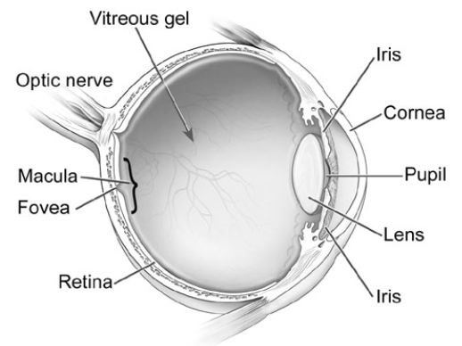
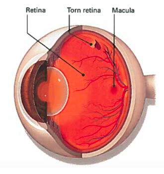
The retina is a layer of nerve tissue that lines the inside of your eye. It consists of light sensitive cells that send signals to your brain and allow for you to see. The retina is very thin, and a tear in it is a very serious and potentially blinding problem. If you develop a retinal tear, it can allow for fluid to enter beneath the retina and cause a retinal detachment. Common symptoms of a retinal tear include the sensation of flashes of light in the eye and floaters. Sometimes a retinal tear can be associated with bleeding into the eye leading to hundreds of new floaters and/or a loss in vision if blood fills the eye.

*Image courtesy of the National Eye Institute http://www.nei.nih.gov
Why do I have a Retinal Tear?
Our eye is filled with a gel-like substance called the vitreous. As we get older this gel breaks down and becomes more liquefied. Eventually, usually after the age of 60, this process of liquefaction causes the gel to separate from the back of the eye. This event is called a posterior vitreous detachment (or PVD). A PVD is completely normal and eventually happens to everyone; however, it is also the time when most eyes have the highest risk of developing a retinal tear. This is because the vitreous gel becomes more mobile and the gel can pull and open up a tear in an area of the retina where is it more adherent to it.
There are several risk factors that put some people at a higher risk for developing a retinal tear. These include:
• Myopia (Near sightedness)
• Having areas of thinning in the retina (lattice degeneration)
• Having had cataract or other interocular surgery
• Having a history of trauma to the eye
• Having a history of a retinal tear or detachment in the fellow eye
• Having family members with a history of retinal tears or detachment
Assessment of a Retinal Tear
We are able to detect a retinal tear during an eye examination. Your surgeon will carefully examine your eye to identify any possible retinal tears. He may need to press on your eye to examine your retina fully. If a retinal tear is present, treatment will be performed right away to prevent a retinal detachment.

How is a Retinal Tear Treated?
A retinal tear is treated by creating scar tissue around the tear to prevent fluid from entering the through the tear and detaching the retina. There are two ways we do this:
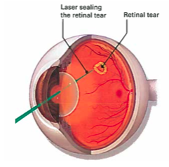
•Laser Photocoagulation: A laser is used to create small burns around the retinal tear. As the burns heal a scar is created around the tear that seals it down. This is the most common way we treat retinal tears.

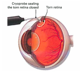
•Cryopexy: Sometimes blood is associated with a retinal tear impeding a good view of it. In these cases it may be difficult to apply laser around the tear and cryopexy may be performed instead. A cryoprobe is pressed on the outside of the eye overlying the tear. Once activated the probe becomes very cold and causes a “freeze burn” around the tear. The residual scarring seals the tear.

If you have had treatment for a retinal tear, you will be given restrictions after treatment, and be reevaluated 1-2 weeks after treatment. Restrictions will include no sudden jarring or head movements as the scarring around the tear is not full strength for approximately 1 week after treatment. If you have acute symptoms of a lot of new floaters, an increase in flashes or a curtain of vision loss you want to call our office right away. This can be a sign of an additional retinal tear or retinal detachment.
Further Information
If you have any questions or concerns regarding this or any other information please call our office at 614-464-3937.
