Surgery Information
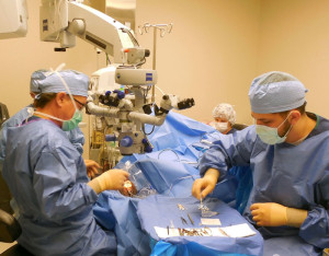
Our surgeons have the privilege of performing their surgical procedures at the Eye Center of Columbus. This is an eye only Ambulatory Surgical Center conveniently located on the 5th floor in the same building as our main office. It is one of the busiest Ophthalmology Surgical Centers in the state of Ohio, performing more than 10,000 surgeries per year. This center prides itself in providing our surgeons with the latest in surgical equipment and technology and our patients with the highest quality surgical care and experience available.
 Our staff will provide you with all necessary instructions in preparation for your surgery. You can expect a phone call from the Surgery Center the day prior to your scheduled procedure. After your surgery, your eye will be taped and shielded, and you will be released after a brief period of observation. You may be instructed to position your head a certain way after surgery. All necessary instructions will be provided to you after your surgery. You will have a postoperative visit scheduled in our office the next day. Bring all prescribed eye drops to this visit so we can clearly provide you with your postoperative instructions.
If you are instructed to position your head after surgery, it is very important to do so or you may risk a suboptimal surgical outcome. We recommend taking no longer then a 5 minute break from positioning every hour.
The four common postoperative surgical positions are:

Given that we perform extremely delicate surgery under a microscope and use technically advanced instrumentation, many of our surgical patients have difficulty envisioning what exactly their surgery will entail. We have provided some surgical videos below that allow you to see some of the common steps and maneuvers we perform during vitrectomy surgery.
Trocar Insertion
– Trocars are ports that allow the surgeon to access the inside of the eye. An infusion line is clipped to one of the ports that infuses fluid into the eye during surgery to keep it formed. The other two ports allow the surgeon to insert instrumentation during surgery.
Core Vitrectomy
– A gel-like substance called the vitreous fills the inside of the eye. During vitrectomy the gel is surgically removed from the eye.
Posteror Vitreous Detachment (PVD) Induction
– The vitreous typically separates spontaneously from the back of the the eye as an eye ages. If it is still attached to the optic nerve during surgery, it is surgically separated. The white substance injected into the eye during the video is Triamcinolone which is often used to better highlight the vitreous.
Epiretinal Membrane (ERM) Peeling
– An epiretinal membrane sometimes forms over the macula causing patients to suffer from distortion and decreased vision. If this occurs, surgery may be indicated to remove the membrane to allow for better vision.
Internal Limiting Membrane (ILM) Peeling
– The ILM is often peeled around a macular hole to help it close. The green substance injected into the eye in the video is a dye that stains the ILM making it easier to identify and peel.
Trocar Removal
– At the end of vitrectomy surgery, the trocars are removed. Often, as in the video, the wounds self seal and no sutures are needed.